


Radiographic examinations are indispensable in modern dentistry, as they assist the dentist in diagnosis, safe treatment planning, and prevention.
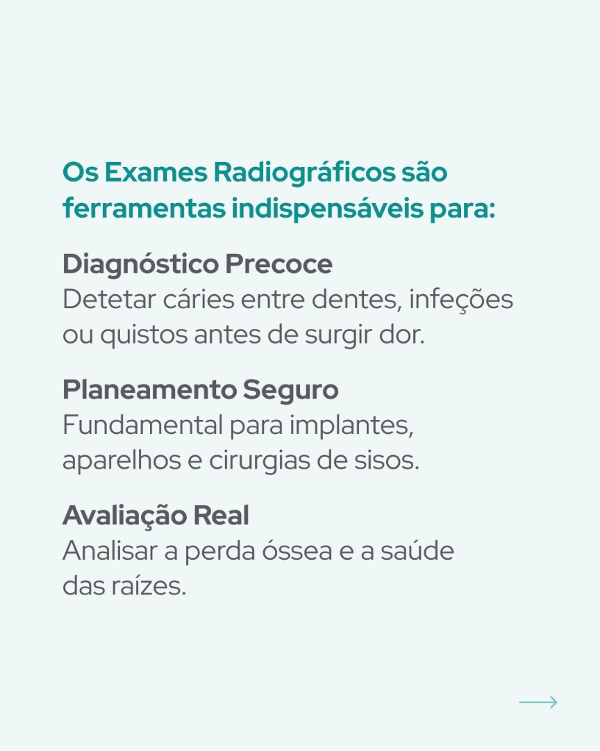
Main Importance and Applications:
- Early diagnosis: Essential for the early detection of interproximal caries (between teeth), bone infections, cysts, and tumours, often before pain symptoms appear;
- Treatment planning: Fundamental in areas such as surgery (impacted wisdom teeth), implantology (assessment of bone volume), endodontics (root canals), and orthodontics (dental alignment);
- Evaluation of structures: Allows analysis of tooth position, periodontal health (bone loss), dental fractures, and temporomandibular joint (TMJ) disorders;
- Monitoring: Used to monitor treatment progress and post-surgical healing.
Most Common Radiographic Examinations:
- Panoramic Radiograph (Orthopantomogram): Provides an overall view of the entire mouth, teeth, and jaws in a single image, ideal for general planning and wisdom tooth assessment;
- Intraoral Radiograph (Periapical/Bitewing): Focuses on specific teeth, showing everything from the crown to the root, ideal for detailed evaluation of caries or root canal treatments;
- Cone Beam Computed Tomography (CBCT): Produces precise 3D images and is widely used in dental implant planning.
The main difference between CBCT (Cone Beam Computed Tomography) and Orthopantomography (panoramic radiography) lies in the dimensionality of the image and diagnostic precision. While orthopantomography provides a two-dimensional (2D), flat image of the entire mouth, CBCT generates detailed three-dimensional (3D) models.
Main Differences Between the Two Examinations:
- Dimensionality and detail: Orthopantomography produces a 2D “stretched” image of the jaws, useful for general check-ups. CBCT, on the other hand, enables 3D visualisation of structures, eliminating image overlap and allowing precise bone measurements;
- Radiation: Orthopantomography uses very low doses of radiation (up to 2 to 3 times lower than CBCT). Although CBCT emits more radiation than panoramic radiography, the levels remain significantly lower than those of a conventional CT scan.
Clinical Applications:
Orthopantomography:
1. Ideal for detecting caries, assessing dental growth in children, and carrying out initial screenings;
2. Provides an overall view of the patient’s oral health, including the number and position of teeth and bone characteristics, among other aspects;
3. Diagnosis of periodontal disease and assessment of the progress of orthodontic treatments;
4. Detection and evaluation of the growth and development of wisdom teeth;
5. Monitoring the development and growth of teeth in children;
6. Diagnosis of sialolithiasis (presence of calculi in the salivary glands);
7. Identification of the location of foreign bodies and detection of lesions such as radicular cysts and other lesions affecting the bony structures of the jaws;
8. Planning of dental extractions and implant placement surgeries;
9. Use in forensic medicine as a method of age estimation.
CBCT:
Indispensable for implant planning, surgery involving impacted teeth (such as wisdom teeth close to nerves), complex endodontic studies, supernumerary teeth, dentoalveolar positional alterations, assessment of lesions in relation to neighbouring anatomical structures, and TMJ pathologies.
Content developed by Dr Rui Salgado, Dental Surgeon
19, May 2026